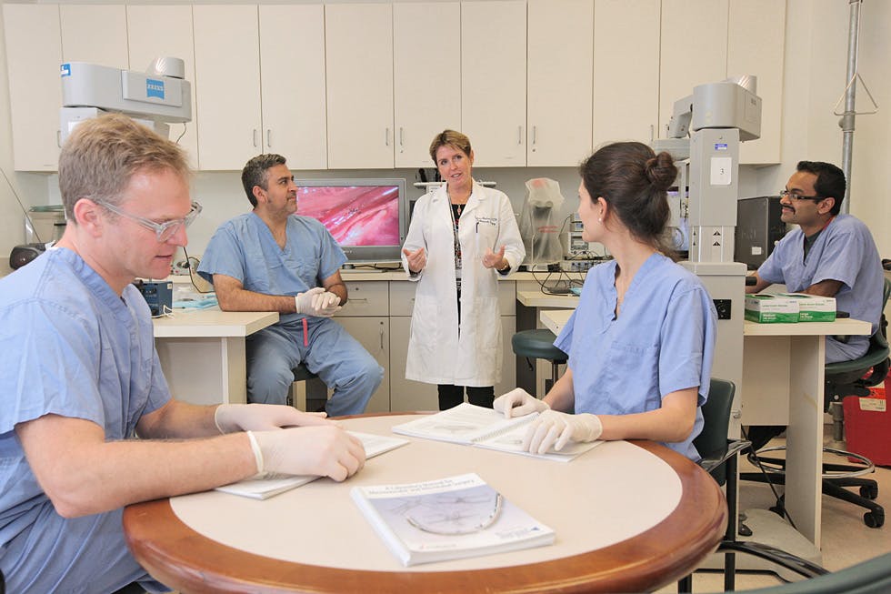
Today, microsurgery is a unique surgical skill that has become a fundamental part of reconstructive surgery and continues to be integrated across many surgical fields. It is widely believed that training in basic microsurgical techniques could help advance the quality of surgical education and ultimately improve patient care.
Invivox met with Dr. Yelena Akelina, a passionate training instructor in practice since 1996 from the Microsurgery Training and Research laboratory at the Department of Orthopaedic Surgery of New York Presbyterian Hospital at Columbia University Irving Medical Center.
INVIVOX: What is microsurgery?
Microsurgery is a surgery performed with operating microscopes, micro-instruments, and micro-sutures ranging from 8.0 to 12.0. Working under high magnification provides the clear visualization needed to repair small vessels and nerves up to 1mm in diameter. This challenging and demanding surgical skill requires careful, professional instruction and a lot of practice. Ideally, microsurgery is taught in a specially designed clinical simulation laboratory equipped with operating room quality surgical microscopes, instrumentation, and sutures, as well as both non-animated and animated models, which include different plastic materials, chicken thighs, and live rats.
INVIVOX: How does microsurgery fit in with other specialties?
Microsurgery is used mostly in hand, plastic, head and neck, and maxillofacial surgery, but the skills also apply to vascular, neurosurgery, transplant surgery and many more. Some specific examples where microsurgery is used include reattachment and revascularization of amputated body parts (replantation), and transplantation of tissue from one part of the body to another (free flap transfer). However, we genuinely believe that the basic principles of microsurgery are beneficial for any surgical specialty.
"After you learn microsurgery, you become a better surgeon."
INVIVOX: What training do you offer?
We offer two courses: Basic Microsurgery and Advanced Microsurgery almost every week of the year. Both classes involve 40 hours of hands-on instruction, completed in one week (Monday through Friday). Our Basic course involves a structured curriculum that consists of gaining working knowledge of the operating room microscope and the anastomoses performed on femoral vessels and sciatic nerves of the rat, 1-mm in diameter. Each exercise starts with the careful dissection of the vessels. Magnification is used every step of the way to ensure careful handling of the delicate tissues. We teach students to use different suturing techniques to complete end-to-end and end- to-side anastomoses, interpositional vein grafts, and epineurial repairs of the sciatic nerve. Our students are gradually moving from arteries to veins and then to grafts and nerves to increase the difficulties of performing more complex procedures. This progression develops an appreciation for the various microsurgical techniques as well as comprehensive planning the steps of each more strenuous exercise.
You can find Dr. Yelena Akelina's courses on Invivox here.
Our Advanced course involves more intricate procedures for more skilled surgeons who had completed the Basic courses or had proven to have sufficient experience. During the Advanced course, they complete similar vascular exercises by scaling down to the epigastric vessels (0.3-0.8 mm diameter), we also perform anastomoses on the carotid vessels, which is more difficult to access due to their anatomical position. Surgeons are also performing different complex free tissue transfers, organs transplants, nerve grafts, and other more complicated procedures.
We very often tailor our exercises to the specialty of the surgeon—we teach organ transplants for transplant surgeons, vasovasostomy for urologists, different free tissue transfers for plastic and reconstructive surgeons, and more.
INVIVOX: Who could come to your training?
There are no prerequisites or special skills needed to participate in our training programs. Anyone can benefit from our intense curriculum–from the student preparing to research animal models, to the resident or fellow completing their training, and the attending surgeon looking to refine one’s technique or expand one’s expertise. We train more than 130 surgeons a year from more than 10 specialties and more than 60 countries.
INVIVOX : Why is it important to learn in the lab? What makes your training different?
We feel that training for such a demanding and difficult skill in the laboratory environment provides many benefits:
- it’s a less stressful atmosphere for learning a challenging hands-on surgical skill
- Students learn under direct supervision-one-on-one training method- and every exercise is evaluated and critiqued by the experienced instructor
- Students are less afraid of making mistakes and instead can learn why they made them and more importantly how to fix them
- Procedures are repeated until they are perfected
- Trainees become much more confident in the operating room and more successful in their procedures, which leads to better clinical outcomes
There are many training courses in microsurgery around the world and most of them are teaching a very similar curriculums but we feel that our program is different and unique because of the following points:
- Hand-on and procedure-structured intensive course
- Individually tailored teaching
- 2:1 student-instructor ratio
- Use of both non-living and live models
- Continuous direct supervision, monitoring, and evaluation of performance
- Using our own training videos as learning tools
- Utilizing the operating room flow meter system as an objective quality assessment tool
- The visual monitoring system in the lab allows us to follow the progress of each student independently and remotely without stressing students
INVIVOX: What are the main benefits of this training? What are the key points attendees can expect to acquire?
The primary goal of this training program is to offer the acquisition of mental and technical surgical skills required to perform delicate procedures. Such skills include:
- Proficiency using the operating microscope at varying levels of magnification
- Gentle handling and dissection of the fragile vascular and nervous tissues
- Eyes-hand-foot coordination
- Attention to small details and planning the steps of the surgery
- Maintaining patience and self-control
- Objective self-assessment of performance
During the course, students learn to complete arterial and venous anastomoses, vein grafts, and nerve repairs. At the beginning of the course, we focus on the quality of the work rather than the speed at which it was completed. With practice comes comfort and speed. By the end of the course, we often expect our students to complete an end-to-end arterial anastomosis within 30 to 40 minutes, compared to the 2.5 hours it takes them at the beginning of the course.
We make sure that the students are not only performing each of the required procedures but also that their anastomoses are patent. We set our standards very high in our course—every procedure is evaluated for patency and needs to be completed with excellence. This type of expectation can only benefit the surgeons in their clinical practice, where the quality of their work will have a direct effect on the outcome of their patients.
INVIVOX: How is Invivox helping you?
Invivox is very important for us because it has a modern and easy to use platform. We can post relevant information to our course as well as, pictures, articles, etc. in an organized and accessible manner for others to see online. People can directly ask questions that Invivox will answer for me and post their feedback about the course. Invivox also takes care of registrations, payments, and travel planning. Before, I had to do all of that by myself.
"I always wanted to improve the social media and Internet presence of our microsurgery courses, but I never felt that I was doing enough. Invivox really helped us to bring it to a more sophisticated level."
INVIVOX: Why is practical training important in your specialty? What does the future hold?
Our future work ultimately depends on clinical needs. When surgeons develop novel techniques, they need new training. For example, we are incorporating new training exercises migrating towards super microsurgery with procedures that would be done with vessels ranging from 0.3mm to 0.8mm. It is a big technical difference and needed to be taught with more meticulousness.
We also hope to include some training in robotic microsurgery in some time in a future. With the development of new and precise machines, robotic microsurgery can significantly reduce hands tremor, morbidity for the patients and improve surgical quality, and so we believe it will be an essential skill for surgeons to learn in the future.
Thank you to my interns Celina Nicolas, John Corvi, YuanDian Zheng for helping with this project.
* Yelena Akelina, DVM, MS is a Research Scientist and a Director/Instructor in Clinical Microsurgery at the Microsurgery Research and Training lab at the Department of Orthopaedic Surgery, Columbia University (New York).

