
INVIVOX: How did autofluorescence imaging come to France?
Dr. Fares BENMILOUD: Autofluorescence (that is to say, the fact that the tissues spontaneously emit fluorescence at certain wavelengths) is known since the 1950s. The discovery of the autofluorescence characteristics of the parathyroid glands is itself fairly recent (2008), and was made at Vanderbilt University, in the United States. Parathyroid autofluorescence imaging was performed for the first time in 2015, in France, at the Gustave Roussy Institute (IGR, Villejuif, France) under the leadership of Dr. Dana Hartl, using the technology of Grenoble-based manufacturer FLUOPTICS© (https://fluoptics.com).
INVIVOX: How has this medical imaging technique improved the management of patients with thyroid and parathyroid disease?
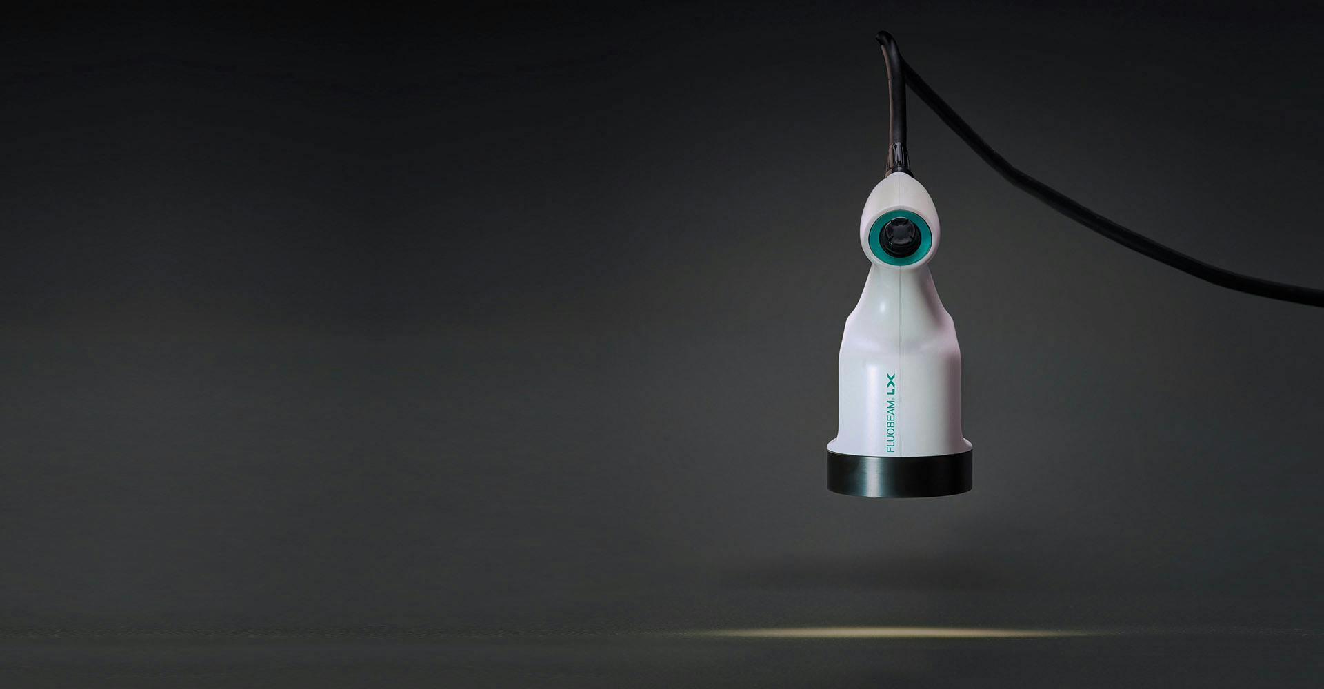
Dr. F. B.: The FLUOBEAM ® LX allows to visualise, without the injection of a contrast agent, the normal parathyroids other than with the eye of the surgeon, and gives the possibility to better preserve them. The method is precise, non-invasive, allowing visualisation in real time. Parathyroids are responsible for regulating the blood calcium level. When we remove a thyroid, we can disrupt their functioning, which requires supplementation with calcium and vitamin D, sometimes for life. This long-term intake is sometimes accompanied by serious treatment-related complications. Parathyroid glands are so small that it is possible to not see them and so to devascularise them by removing the thyroid gland. To be able to view them upstream allows the surgeon to be more conservative.
INVIVOX: Can you describe the procedure in detail?
Dr. F. B.: There are two phases: first, the autofluorescence exploration, that is the fact that the parathyroids spontaneously emit a natural fluorescent signal when they are stimulated with a certain light, and the injection of a fluorescent dye, the indocyanine green, which allows to visualise the vessels, and then, at the end of the procedure, a second injection which allows to observe if the parathyroids remain alive after the operation. We use the FLUOBEAM ® LX which is a camera linked to a panel PC and whose controls are activated by the surgeon via a joystick to increase sensitivity, to zoom etc.
"Autofluorescence is a disruptive technology. Previously, there was no examination available to see normal parathyroids during the operation and without contrast agent."
INVIVOX: How do you explain the decrease of hypocalcaemia cases?
Dr. F. B.: I think that the decrease of hypocalcaemia cases is related to the fact that we operate better thanks to this machine which allows us to see the parathyroids very early in the procedure. It allows us to refocus on parathyroid dissection. Several recent studies show that definitive hypocalcaemia increases mortality. With this technique, I think we will be able to dramatically reduce complications. Since using autofluorescence, I have had no case of definitive hypocalcaemia and about 5% transient hypocalcaemia. By using vascular mapping through the injection of indocyanine green, the rates are even lower: in one year, I did not have a single case of hypocalcaemia. The current rates observed in publications are 6 to 10% of definitive hypocalcaemia and 30 to 40% of transient hypocalcaemia.
INVIVOX: Does it also facilitate parathyroidectomy procedures?
Dr. F. B.: Yes, it may speed up parathyroid exploration or guide resection in complex cases, but the main interest of this technique, for me, lies in the limitation of parathyroid risks during thyroidectomy. For parathyroidectomies, we already have very effective preoperative exams that allow us to detect pathological parathyroidisms to remove them.
INVIVOX: How do your fellow endocrinologists welcome this technique? Do many of you know how to use it?
Dr. F. B.: As this technology is relatively new, the need for training is relatively high. We are only a few who use and master this embedded imaging technique. As with any innovation, there are currently some obstacles like not having enough scientific proof of its efficiency and justification for its use. The first version of the FLUOBEAM ® was less ergonomic with a lower quality image than the FLUOBEAM ®LX just released. For my part, in view of the studies that I made and that I will publish, as for example in November in the JAMA Surgery (https://jamanetwork.com/journals/jamasurgery) and present orally at the congress of the American Thyroid Association (https://www.thyroid.org/) which takes place at the end of October in Chicago, I am convinced of its effectiveness.
INVIVOX: Will parathyroid fluorescence imaging become a standard in thyroid and parathyroid surgery?
Dr. F. B.: Yes, in my opinion. It is already part of my routine and will probably soon be that of all thyroid surgeons. It is also important to point out that this is a tool, and as such, in no case should it be a substitute for the surgeon's judgment: once we have seen the parathyroids, we must fight to save them. The results are operator dependent.